June 14, 2015 - A new engineered positron-emitting imaging agent that targets prostate-specific membrane antigen (PSMA) can detect metastatic prostate disease better than conventional imaging, according to a study1 presented at the Society of Nuclear Medicine and Molecular Imaging (SNMMI) 2015 Annual Meeting.2
The engineered radiotracer "can assist in guiding biopsies to improve yield and can help identify occult sites of disease,” said one of the authors of the study, Neeta Pandit-Taskar, MD, from Memorial Sloane Kettering Cancer Center in New York City, during a press conference at the SNMMI annual meeting. She added that it “offers the potential for direct imaging of metastatic prostate cancer."
The minibody (IAB2M) is labeled with radioactive zirconium-89 to create a radiotracer known as Zr-89 Df-IAB2M, and is used with positron emission tomography–computed tomography (PET/CT).
The can detect the prostate cancer cells that have metastasized to bone — one of the most difficult areas to evaluate using standard methods,” said Dr. Pandit-Taskar.
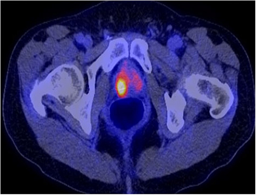
Image: 18F-FDG PET/CT shows intense hypermetabolism in right prostate lobe. Source: http://jnm.snmjournals.org/content/52/1/81/F1.expansion.html
In the Phase I study, the researchers evaluated 393 patients with metastatic prostate lesions using computed tomography (CT), magnetic resonance imaging, molecular bone scans, fluorodeoxyglucose radiotracer positron emission tomography (FDG-PET) and the new agent Zr-89 IAB2M PET/CT (IAB2M PET/CT). Researchers identified 25% more lesions than other imaging modalities using Zr-89 Df-IAB2M PET/CT imaging, detected 82% of bone lesions compared with 57.7% detected by bone scans, 53% by CT, and 26.2% by FDG-PET. Zr-89 Df-IAB2M PET/CT imaging also detected 86% of soft tissue lesions compared with the 65.8% identified with CT and the 48.2% identified with FDG.
The results showed 65 bone and 32 soft tissue sites were visualized exclusively with minibody imaging. Based on the data, the authors concluded that 89Zr IAB2M Mb imaging is safe, has favorable biodistribution and kinetics with possible detection of lesions at 24 - 48 h p.i. Imaging with higher Mb mass is planned.
Source:
- Pandit-Taskar N, et al. 89Zr-IAB2M minibody imaging in patients with prostate cancer: Biodistribution, kinetics, lesion uptake and organ dosimetry. Society of Nuclear Medicine and Molecular Imaging (SNMMI) 2015 Annual Meeting: Abstract 400. Presented June 9, 2015.


