March 16, 2016 - A new, real–time method of imaging molecular events after strokes uses cell-penetrating peptides in combination with magnetic resonance imaging (MRI) to detect and track gelatinase (MMP-2/-9) activity, according to a recent study1 published in the Journal of Cerebral Blood Flow and Metabolism.
Matrix metalloproteinases (MMPs), particularly gelatinases (MMP-2/-9), are involved in neurovascular impairment after stroke. Detection of gelatinase activity in vivo can provide insight into blood-brain barrier disruption, hemorrhage, and nerve cell injury or death. Researchers applied gelatinase-activatable cell-penetrating peptides (ACPP) with a cleavable l-amino acid linker to examine gelatinase activity in primary neurons in culture and ischemic mouse brain in vivo. Fluorescence intensity was significantly reduced when cells or mice were treated with MMP inhibitors or when a cleavage-resistant ACPP-Cy5 was substituted. We also applied an ACPP dendrimer (ACPPD) conjugated with multiple Cy5 and/or gadolinium moieties for fluorescence and magnetic resonance imaging (MRI) in intact animals. Fluorescence analysis showed that ACPPD was detected in sub-femtomole range in ischemic tissues. Moreover, MRI and inductively coupled plasma mass spectrometry revealed that ACPPD produced quantitative measures of gelatinase activity in the ischemic region. The resulting spatial pattern of gelatinase activity and neurodegeneration were very similar. We conclude that ACPPs are capable of tracing spatiotemporal gelatinase activity in vivo, and will therefore be useful in elucidating mechanisms of gelatinase-mediated neurodegeneration after stroke.
"During an ischemic stroke, harmful enzymes called gelatinase become overactive in areas of the brain where blood flow is cut off, said Zezong Gu, PhD, an associate professor of pathology and anatomical sciences at the MU School of Medicine and lead author of the study. "Over-activation of these enzymes causes brain damage. Our team hypothesized that if we could visualize and track this activity in real-time, we could then work on developing a way to block the activity and prevent brain damage from occurring."
"Our findings indicate that tagged peptides can be used as a non–invasive probe to detect and track gelatinase activity," Gu said. "This process may serve as an additional tool for clinicians to treat their patients if a viable inhibitor can be developed to prevent the damage caused by this activity."
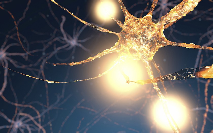
Figure. Real-time imaging of neurological damage could lead to improved stroke care.2
References:
1. Chen S1, Cui J2, Jiang T3, Olson ES3, Cai QY4, Yang M4, Wu W1, Guthrie JM5, Robertson JD5, Lipton SA6, Ma L4, Tsien RY7, Gu Z8. Gelatinase activity imaged by activatable cell-penetrating peptides in cell-based and in vivo models of stroke. J Cereb Blood Flow Metab. 2015 Dec 17. pii: 0271678X15621573.
2. New Imaging Technique May Give Physicians Clearer Picture of Stroke Damage. MU School of Medicine. March 11, 2016. http://medicine.missouri.edu/news/20160311-new-imaging-technique.php


